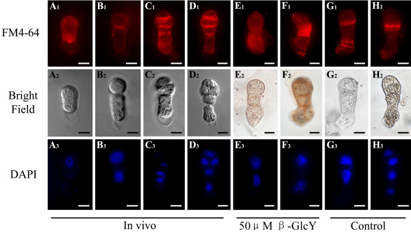
Figure 7 .
Localization of endocytic vesicles by FM4-64 staining in tobacco zygotes and proembryos. (A1-H1) The fluorescence images of living zygotes and proembryos stained by FM4-64. In a zygote, the fluorescence of FM4-64 evenly distributes in the whole cytoplasm (A1). During cell plate formation, endocytic vesicles mainly localize in the new cell plate edges (B1-D1). After treating with 50 μM β-GlcY, the fluorescence of FM4-64 evenly distributes in cytoplasm of in vitro cultured proembryos (E1 and F1). In proembryos cultured without β-GlcY, the fluorescence images are similar to that of in vivo proembryos (G1). (A2-H2) Bright-field images of zygotes and proembryos corresponding to A1-G1. (A3-H3) Fluorescence images of DAPI-stained nuclei corresponding to bright-field images. Bars = 10 μm.

