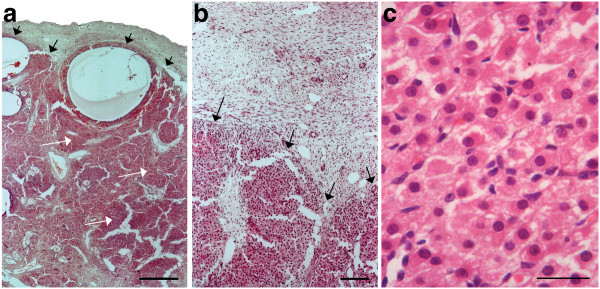
Figure 3.
Sections of the ovaries of an elephant fetus at 13.5 months of gestation.a) The black arrows indicate the cortico-medullary border. The white arrows indicate interstitial tissue (Scale bar = 700 μm). b) The border between cortical tissue at the top of the photograph and interstitial tissue at the bottom (Scale bar = 350 μm). c) High power magnification of the interstitial cells shown in b (Scale bar = 20 μm).

