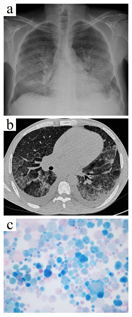
Figure 4.

Case 3. The chest radiograph (a) showed diffuse nonspecific alveolar infiltrates. Chest computed tomography (b) revealed areas of ground-glass attenuation interspersed with normal areas, as well as a bilateral pleural effusion. The bronchioloalveolar lavage showed many blue hemosiderin-laden macrophages consistent with diffuse alveolar hemorrhage (c, Perls’ stain, x400).
