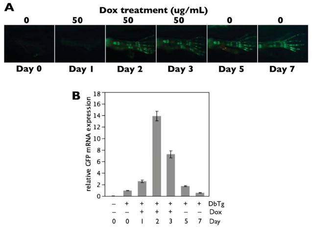
Figure 4.
GFP mRNA and protein stability in transgenic animals from the rtTA-dependent system. A) Double transgenic tadpoles were treated with 50μg/mL Dox for 3 days at NF57. Green fluorescence images of the hind limbs were taken on Days 0–3, 5 and 7. GFP protein became strongly visible starting on Day 2 and remained intense through Day 7—four days after Dox removal. B) GFP mRNA was measured using quantitative real-time PCR from hind limbs corresponding to the images in the previous panel, n = 3 pairs of limbs per day. Minimal GFP mRNA was detectable in transgenic animals in the absence of Dox, and GFP mRNA expression reached a peak at 2–3 days of Dox treatment. By 2–4 days after Dox removal, GFP mRNA quickly returned to pre-Dox levels. Note that the persistence of inducible GFP protein expression, after GFP mRNA has degraded, renders this system useful for labeling and tracking tadpole cartilages through metamorphosis.

