FIGURE 4.
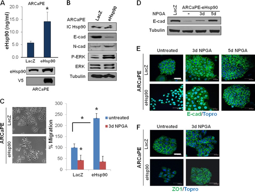
Modest elevation of eHsp90 is sufficient to suppress E-cadherin function and promote cell motility. A, upper panel, ELISA analysis of secreted eHsp90 protein detected from conditioned media collected from parental ARCaPE cells stably transduced with control (lacZ) or V5-tagged eHsp90α lentivirus. Lower panel, immunoblot detection of total (endogenous and exogenous) eHsp90α, or V5 detection of transduced eHsp90 protein. B, immunoblot analysis of cell lysates from ARCaPE-LacZ or ARCaPE-eHsp90 confirmed consistent levels of intracellular eHsp90α (IC Hsp90). Indicated analysis of E- and N-cadherin and ERK activity. C, representative morphology of the indicated ARCaPE cells. Analysis of cell motility of ARCaPE-eHsp90 either untreated or treated with NPGA. D, effect of NPGA upon E-cadherin expression in ARCaPE-eHsp90. E, analysis of E-cadherin localization in ARCaPE-LacZ and ARCaPE-eHsp90 untreated cells, or treated for the indicated times with NPGA. F, membrane localization of ZO1 in ARCaPE-LacZ and ARCaPE-eHsp90 untreated cells, or treated with NPGA for 3 days. Asterisks (*) indicate significance of p value ≤0.05. Scale bar is 50 μm.

