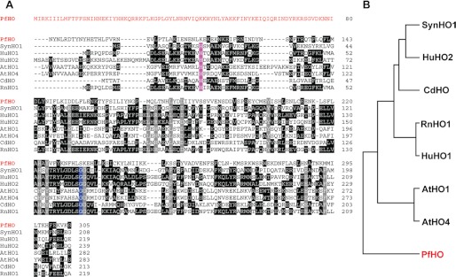
FIGURE 5.
Sequence analysis of PfHO relative to known heme oxygenases. A, ClustalW alignment of PfHO with known heme oxygenases from Synechocystis sp. 6803 (SynHO1), H. sapiens (HuHO1 and HuHO2), A. thaliana (AtHO1 and AtHO4), Corynebacterium diphtheriae (CdHO), and R. norvegicus (RnHO1). The amino acid leader sequence of PfHO is shown in red. Conserved residues in all seven known HOs are colored gray. The conserved proximal His residue that coordinates the bound heme and the conserved distal Gly residue are colored violet and blue, respectively. Identical residues in three or more proteins are in black. For simplicity, the chloroplast-targeting leader sequences of AtHO1 and AtHO4 and sequences of all proteins that extend beyond the C terminus of PfHO have been omitted. B, phylogenetic relationship of PfHO relative to the aligned heme oxygenases.

