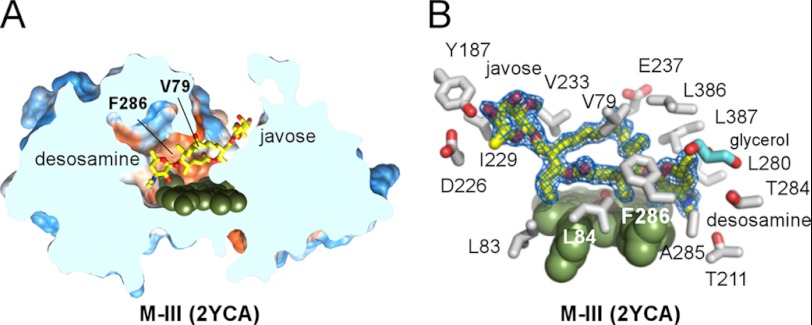
FIGURE 5.
Parallel “desosamine-in javose-out” M-III-binding mode. A, slice through the parallel to the heme-binding site accommodating M-III. B, M-III is well defined by the 2Fo − Fc electron density map contoured at 1.0 σ in the 2YCA structure (blue mesh). Selected side chains within 5 Å of the M-III are shown in gray. The glycerol molecule (cyan) is bound in the vicinity of desosamine, partially filling the void space of the buried cavity.

