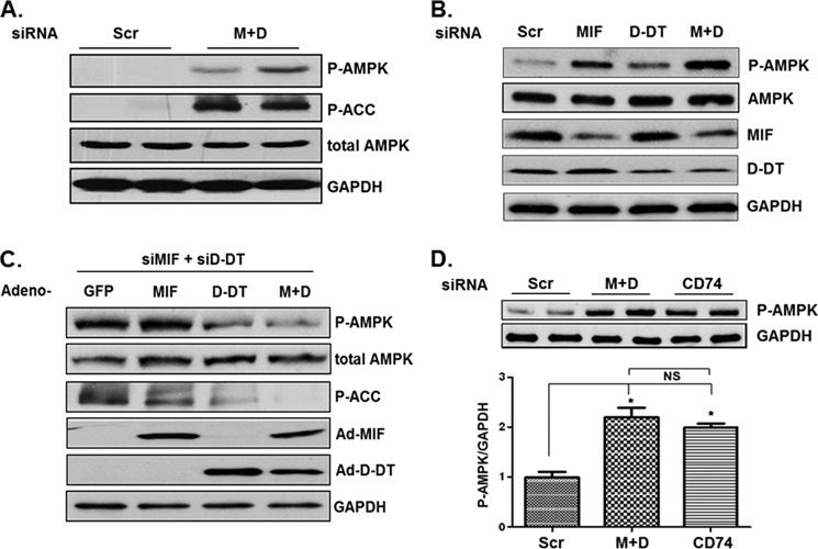
FIGURE 1.
MIF and d-DT cooperate to antagonize AMPK activation. Scrambled (Scr), MIF, and/or d-DT siRNAs were transfected into A549 cells using Oligofectamine as indicated for 72 h (A and B), and lysates were examined by immunoblotting. C, A549 cells were siRNA-transfected as indicated for 48 h and then infected with GFP (used as a control for adenoviral infection/overexpression), MIF, d-DT, or MIF + d-DT (M+D) adenovirus overnight. Lysates were examined by immunoblotting. D, A549 cells were siRNA-transfected as indicated for 72 h, and lysates were examined by immunoblotting. Bio-Rad Quantity One software was used for densitometry and P-AMPK/GAPDH (top panels, n = 2) densitometry values are depicted in the graph. *, p < 0.05 by one-way ANOVA analysis is shown for individual group comparisons. Data shown are representative of two independent experiments. Error bars, S.E. MIF, d-DT, and CD74 were reduced by ∼85%, 70%, and 80%, respectively (not shown).

