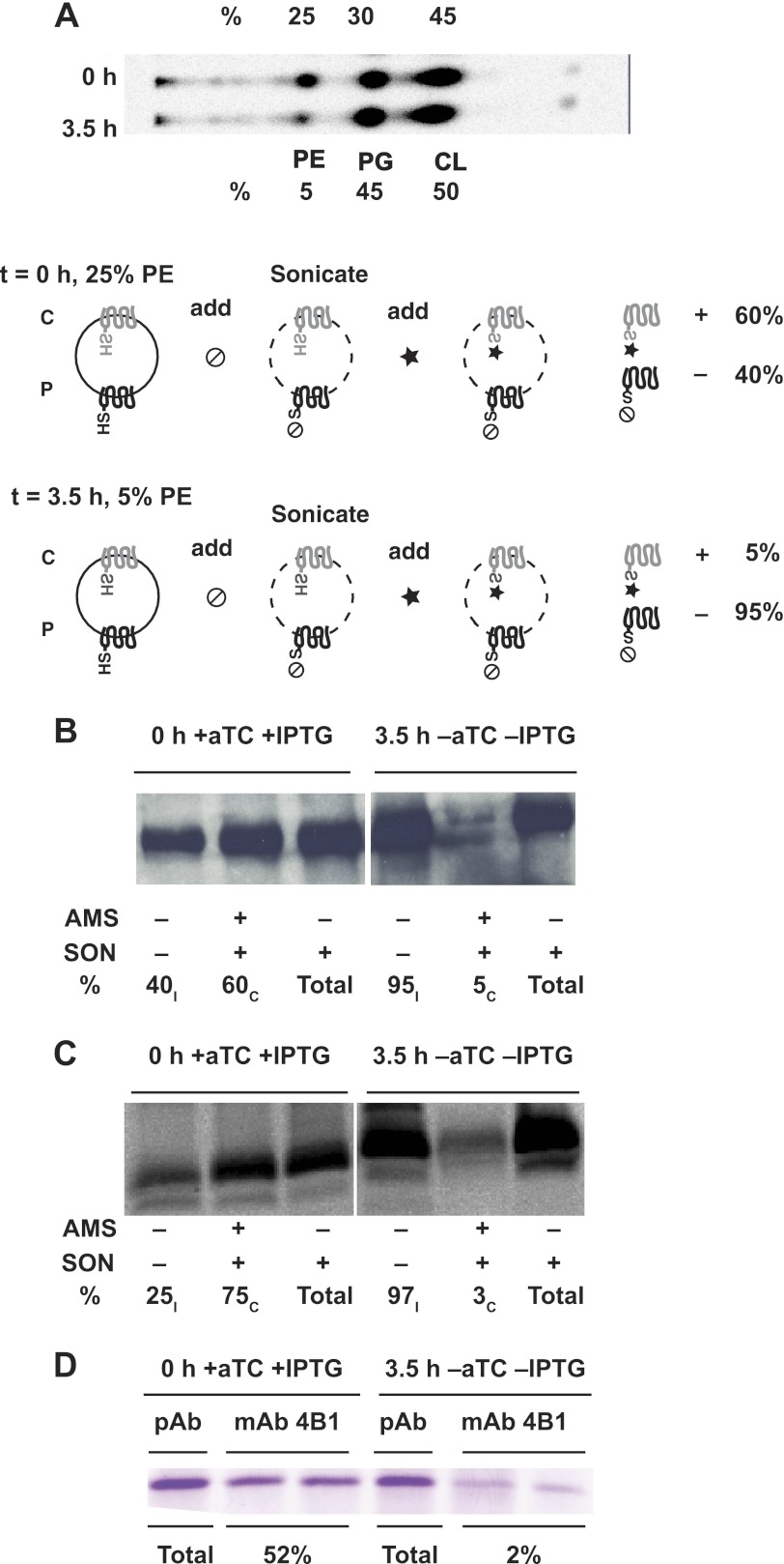
FIGURE 5.
Elimination of PE triggers post-insertional inversion of LacY. Cells expressing LacY with either a H205C replacement in C6 or a L14C replacement in N-terminal were cultivated first in the presence of 100 ng/ml of aTc to adjust PE content to an intermediate level. Cells were switched to growth for 3.5 h without IPTG and aTc to deplete PE in the absence of newly synthesized LacY. A, phospholipids were radiolabeled ([32P]PO4) during growth with IPTG and 100 ng/ml of aTc (top lane). Cells were switched to growth in the absence of IPTG and aTc but in the presence of [32P]PO4 for 3.5 h (bottom lane). Phospholipid composition expressed as mole % was determined using solvent system 2 as described under “Experimental Procedures.” B and C, intact whole cells grown in parallel without [32P]PO4 were treated with MPB as described under “Experimental Procedures” without sonication (SON, −), prelabeled (+) or not (−) with AMS, or after sonication (SON, +). Labeling was performed for H205C (C6, B) and L14C (NT, C) replacements, either after initial assembly of LacY in cells containing ∼25% PE (0 h + aTc + IPTG) or after cessation of PE and LacY synthesis for 3.5 h (3.5 h −aTc, −IPTG). The ratio of inverted (subscript I) and correct (subscript C) was calculated from the intensity of biotinylated bands by using Quantity One software for scanning and analysis of the captured images (Bio-Rad). Each lane represents aliquots of an equal volume of cell culture. D, cells were grown and harvested as indicated in B and C. Aliquots of equal volumes of cell culture were subjected to immunoprecipitation by pAb (total LacY) or mAb 4B1 (properly oriented LacY) as described under “Experimental Procedures.” Gel bands at the position of LacY after SDS-PAGE were quantified (mAb 4B1) and expressed relative to the band at the far left (pAb). The two bands under mAb4B1 represent duplicate samples.

