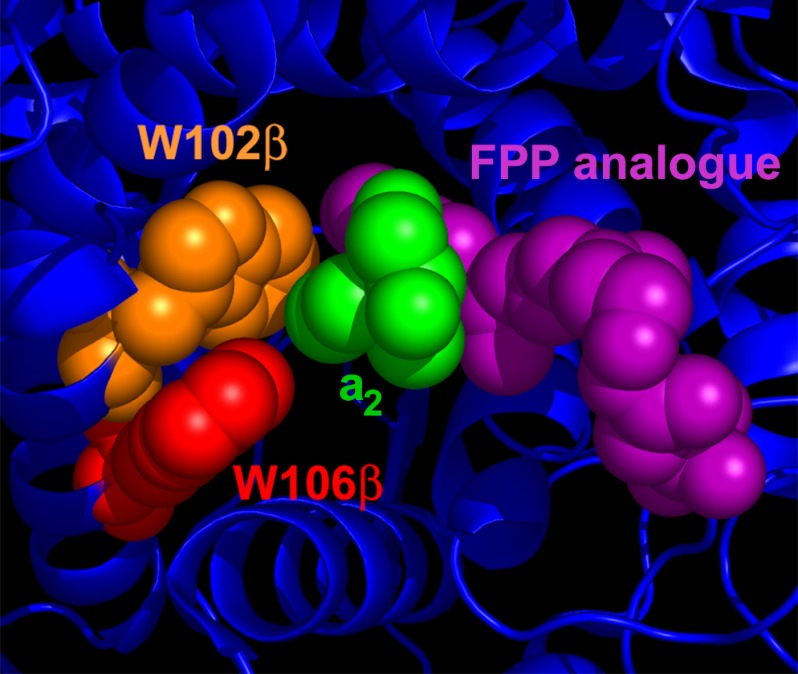
FIGURE 1.
FTase active site structure. Structure of a peptide substrate bound to FTase illustrate the A2 residue binding pocket. The A2 residue of the peptide substrate KKKSKTKCVIM (green) is surrounded by residues Trp-102β (orange), Trp-106β (red), and Tyr-361β (not shown) within the active site of FTase. The A2 residue also contacts the isoprenoid tail of the FPP analog inhibitor FPT-II (purple). The figure was derived from PDB ID 1D8D and adapted from Reid et al. (9).

