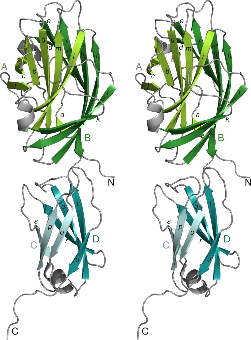
FIGURE 3.
Structure of the N-terminal domain of Epf (EpfN), shown in stereo. The β-sheets of the EpfN1 subdomain are in green (sheet A lighter, sheet B darker), and those of the EpfN2 subdomain are in blue (sheet C lighter, sheet D darker). Lowercase letters indicate β-strands, and uppercase letters indicate the β-sheets and the N/C chain termini.

