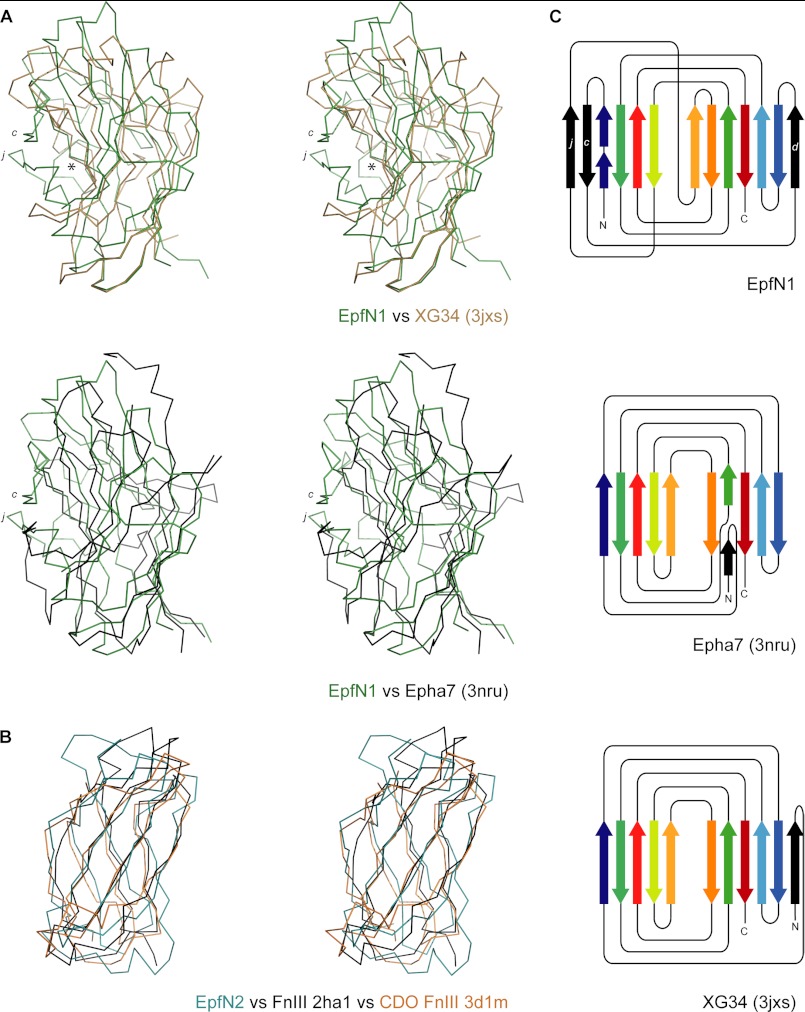
FIGURE 4.
Structural relationships of the EpfN subdomains EpfN1 and EpfN2. A, structural superposition of EpfN1, in green, on to the carbohydrate-binding module of XG34 (PDB code 3jxs) (49), in brown, and the ligand-binding domain of the human ephrin receptor Epha7 (PDB code 3nru), in black; c and j designate the additional β-strands in EpfN1, and the asterisk indicates the carbohydrate-binding site in XG34. B, structural superposition of EpfN2, in green, on to the fibronection type III domains of fibronectin (2ha1) (43), in black, and human CDO (3d1m) (50), in brown. In each case, the fold is represented as a Cα trace in stereo; structures were aligned using SSM (31). C, topology diagrams of EpfN1, Epha7, and XG34. Aligned β-strands are marked in the same colors, and additional β-strands are colored black. For clarity, decorating α-helices, which are present in all three structures, were omitted, and the lengths of β-strands are not to scale.

