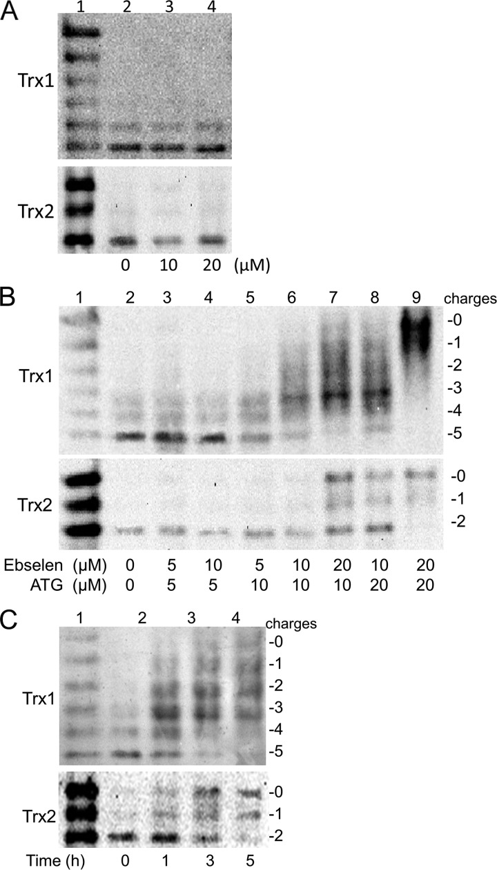
FIGURE 5.
The Redox state of Trx1/2 in HeLa cells exposed to ATG and ebselen. A, HeLa cells were treated with the indicated concentration of ebselen (lanes 2-4) for 24 h, and then the redox states of Trx1 and Trx2 in HeLa cells were detected with a redox state Western blot analysis. Lane 1, mobility standards. B, HeLa cells were treated by the indicated concentrations of ATG for 6 h, and then ebselen was added into the medium (lanes 2-9). After 24 h, the redox state of Trx1 and Trx2 in HeLa cells were detected with a redox state Western blot analysis. Lane 1, mobility standards. C, HeLa cells were treated with 20 μm ATG for 6 h (lane 3-5), and then ebselen was added into the medium (lane 3-5). At the indicated time point, the redox states of Trx1 and Trx2 in HeLa cells were detected with a redox state Western blot analysis. Lane 1, mobility standards. Lane 2, HeLa cells without treatment.

