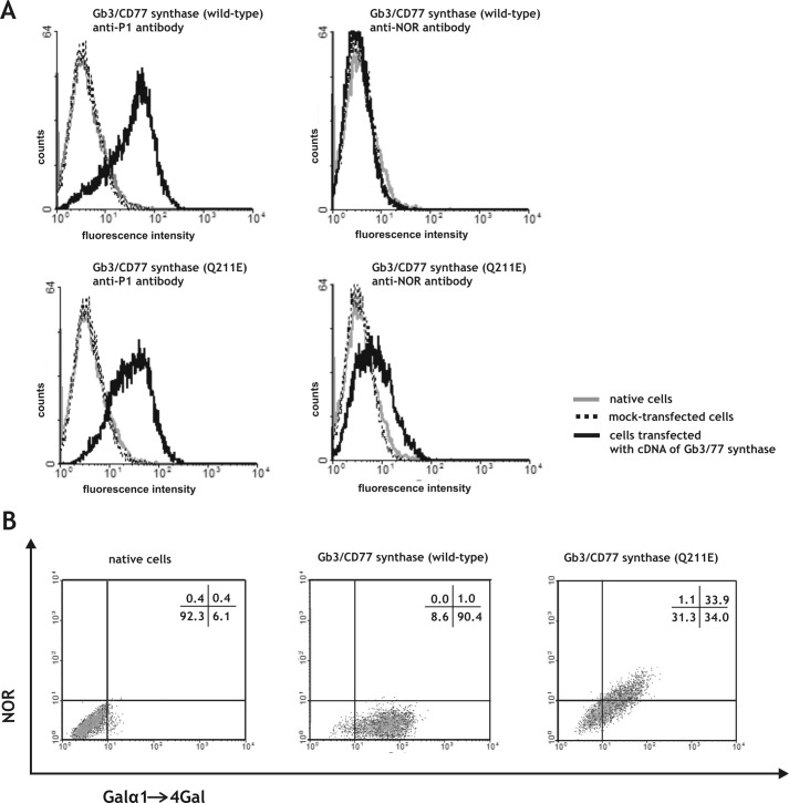
FIGURE 2.
A, flow cytometric analysis of the binding of anti-P1 and anti-NOR antibodies to 2102Ep cells transfected with vectors encoding the consensus Gb3/CD77 synthase (containing a Gln residue at position 211) or Gb3/CD77 synthase containing a Glu at position 211 (Q211E). B, two-dimensional flow cytometric analysis of 2102Ep cells transfected with vectors encoding the consensus Gb3/CD77 or Gb3/CD77 Q211E synthase. The cells were coated with anti-P1 and anti-NOR antibodies. Percentages of cell number in each quadrant are indicated.

