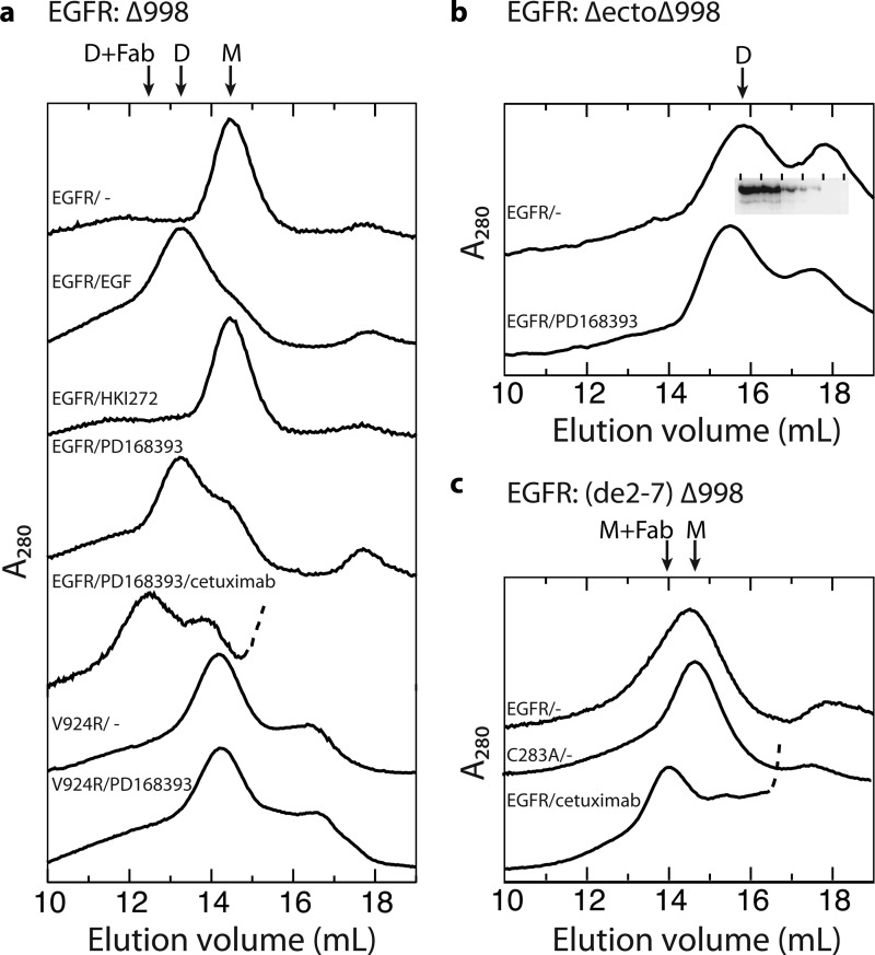
FIGURE 4.
Gel filtration chromatograms of detergent-solubilized, purified EGFR proteins. The purified EGFR truncation mutants indicated in panels a–c, with any additional mutations shown above each trace (0.7 μm), were treated with or without kinase inhibitors (200 μm), EGF (20 μm), or cetuximab Fab (40 μm) for 5 min on ice for 5 min and subjected to gel filtration on a Superose 6 HR column equilibrated with running buffer containing 0.2 mm dodecylmaltoside and 0.2 mm DTT. The inset in b shows Western blots with protein C antibody of fractions from the upper trace, demonstrating that EGFR is present only in the dimer peak, and not in the second peak. The arrows mark positions of dimeric (D), monomeric (M), and Fab complex material (Fab). Traces of Fab complexes are truncated so that only the beginning, rising portion of the Fab peak is shown, which is dashed.

