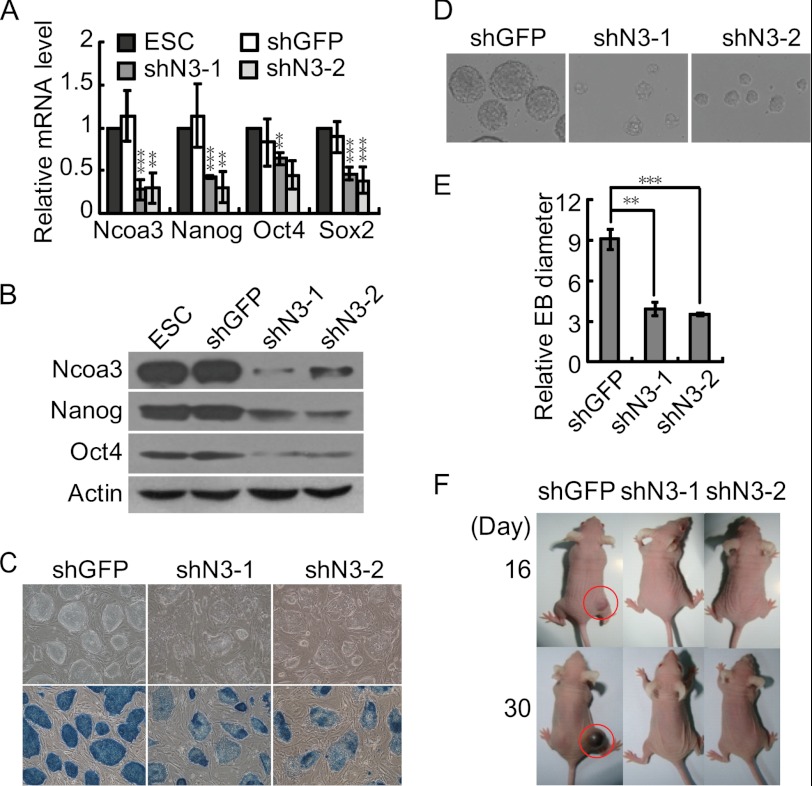
FIGURE 3.
Ncoa3 depletion impairs ESC differentiation in vitro and in vivo. A, reduced expression of pluripotency genes in two stable Ncoa3 knockdown ESC lines. Stable shGFP control and Ncoa3 knockdown ESC lines were established by transfection of shGFP or shRNA plasmids targeting Ncoa3 (shN3-1 and shN3-2) into V6.5 ESCs, followed by puromycin selection. The expression levels of Ncoa3, Nanog, Oct4, and Sox2 in these cell lines were quantified with quantitative RT-PCR. Means ± S.D. from three independent experiments were plotted. B, Western blot analysis showed that the protein levels of Ncoa3, Nanog, and Oct4 in stable Ncoa3 knockdown ESC lines were decreased. β-Actin served as a loading control. C, alkaline phosphatase staining in stable shGFP and Ncoa3 knockdown ESCs. D, phase contrast images of day 2 EBs formed by stable shGFP and Ncoa3 knockdown ESCs. The size of day 2 Ncoa3 knockdown EBs was smaller than that of shGFP control EBs. E, 20 each of randomly selected shGFP and Ncoa3 knockdown EBs were scored, and their relative diameters from images taken under identical magnification were calculated. The means ± S.D. of relative diameters are presented. F, representative pictures of mice at day 16 and day 30 after injection of ESCs. Stable shGFP and Ncoa3 knockdown ESCs were injected subcutaneously into nude mice. Teratoma (red circles) formed by shGFP ESCs were observed as early as 16 days after injection. However, no teratoma was formed from shN3-1 or shN3-2 ESCs up to 30 days. **, p < 0.01; ***, p < 0.001.

