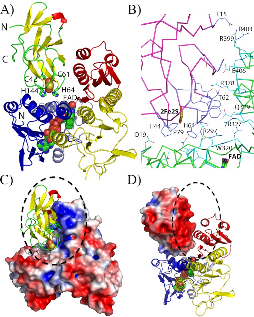
FIGURE 4.
Structure of the reductaseTOL-ferredoxinTOL complex. A, overall structure of the reductaseTOL-ferredoxinTOL complex. The schematic representing ferredoxinTOL is colored according to secondary structure with β-sheets in yellow, α-helices in red, and loop regions in green. Residues coordinating the Rieske-type [2Fe-2S] cluster are shown as sticks and are labeled. ReductaseTOL is color-coded as in Fig. 3A with the FAD-binding domain in blue and light blue, the NADH-binding domain in yellow, and the C-terminal domain in red. B, binding interface between ferredoxinTOL(magenta ribbons; blue carbon atoms) and reductaseTOL(green ribbons; light blue carbon atoms). C, electrostatic potential mapped on the solvent-accessible surface of reductaseTOL with ferredoxinTOL displayed as in A. D, electrostatic potential mapped on the solvent-accessible surface of ferredoxinTOL with a schematic representation of reductaseTOL as in A.

