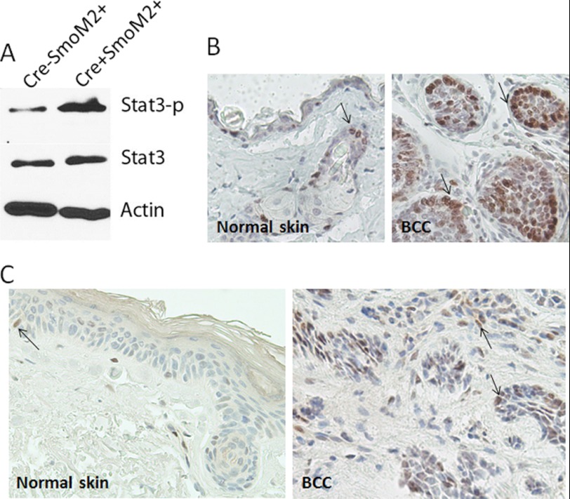
FIGURE 2.
Elevated level of STAT3 phosphorylation in SmoM2-mediated BCCs of mice and humans. Specific antibodies to pSTAT3-Y705 were used to detect STAT3 activation in mouse epidermis by Western blotting (A) and immunohistochemistry (B and C) according to the protocol of the manufacturer. The epidermis was separated from the dermis by merging the skin in dispase® (5 mg/ml in PBS) for 2 h at 37 °C. The protein loading controls were endogenous STAT3 and β-actin. The negative control for immunohistochemistry was one section without primary antibodies. Positive staining is shown in brown (arrows). Tissues were counterstained with hematoxylin (blue). Positive staining of pSTAT3-Y705 was seen in the nucleus.

