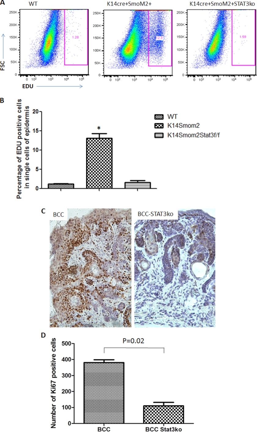
FIGURE 4.
The effect of STAT3 knockout on cell proliferation in SmoM2-expressing epidermis. We compared K14cre+/SmoM2YFP+/STAT3wt mice with K14cre+/SmoM2YFP+STAT3f/f mice for cell proliferation in epidermis by EdU labeling and Ki-67 staining. EdU labeling was performed 14 h before animal sacrifice, and positive cells were analyzed by flow cytometry (A). B, data summary from all mice from each group. The percentage of EdU-positive cells in tumor-bearing K14cre+/SmoM2YFP+/STAT3wt mice (K14creSmom2) compared with that from K14cre+/SmoM2YFP+STAT3f/f mice (K14creSmom2STAT3f/f) (p < 0.0001). Immunohistochemistry was performed with specific antibodies to Ki-67 (C), and the number of Ki-67 positive cells/field (×200, one field = 0.9 mm2) was counted under the microscope. The number of positive cells shown in D was an average from five mice (eight fields per mouse). *, p < 0.05.

