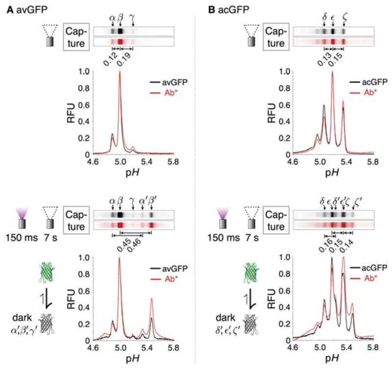
Figure 2.

Probed isoelectric focusing of avGFP and acGFP reveals base-shifted reversibly photobleached isoforms. (A) Static LAVAgel fluorescence images and electropherograms of immobilized avGFP isoforms (bright:α, β, γ; dark: α′ and β′; γ′ is below the assay limit of detection) within the microchannel (black; excitation 445–495 nm, emission 508–583 nm) after focusing under - top: nil light exposure conditions, bottom: 150 ms pre-exposure to 100% UV (270 mW cm−2, 300–380 nm). Isoform capture was initiated immediately after the indicated pre-exposure protocol by 15 s UV irradiation of the LAVAgel under non-focusing conditions. Red fluorescence (excitation 525–555 nm, emission >575 nm) gel images and electropherograms are produced following pH gradient washout and LAVAgel probing with 600 nM Texas Red-labeled anti-GFP antibody (Ab*) for immobilized isoforms. N.B. fluorescence of captured dark isoforms (α′ and β′) is switched on during imaging under blue illumination. (B) The corresponding micrographs and electropherograms for immobilized acGFP isoforms (bright:δ, ε, ζ; dark: δ′, ε′ and ζ′) probed with the same anti-GFP antibody.
