Figure 2.

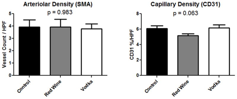
Vessel Density in the left ventricle. Arteriolar density identified by structures of the appropriate size co-staining for smooth muscle actin (SMA) and CD31, whereas capillary structures stain for CD31 alone. HPF = high powered field.

Vessel Density in the left ventricle. Arteriolar density identified by structures of the appropriate size co-staining for smooth muscle actin (SMA) and CD31, whereas capillary structures stain for CD31 alone. HPF = high powered field.