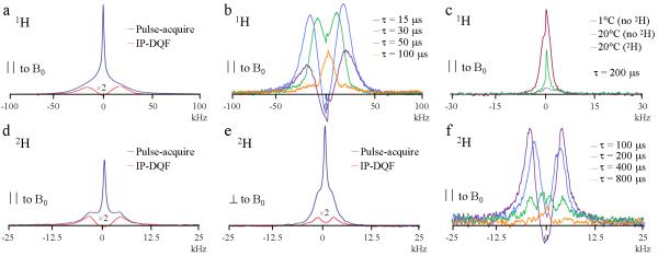
Figure 2.
NMR spectra of an anterior sample of a cortical bone specimen of the mid-shaft tibia from a 37-year old male donor after equilibration in 2H2O recorded at 20°C. All spectra showed similar behavior over a range of temperatures (5-50°C). a) 1H pulse-acquire and IP-DQF (at creation time, τ = 25μs) spectra with the bone’s longitudinal axis aligned parallel (∥) to the magnetic field, B0. b): 1H IP-DQF spectra at various τ. c): 1H IP-DQF spectra (τ = 200μs) collected under various conditions. ‘No 2H’ and ‘2H’ refer to with and without 2H2O immersion. d) and e): 2H pulse-acquire and IP-DQF (τ = 200 μs) spectra with the bone’s longitudinal axis aligned ∥ and perpendicular (⊥) to B0. f): 2H IP-DQF spectra at various τ.

