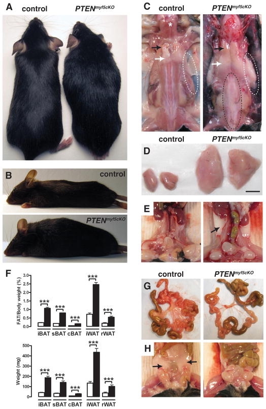
Figure 1. Deleting PTEN in the Myf5+ lineage causes severe combined lipomatosis and partial lipodystrophy.
(A) Anatomy of a 6-week-old PTENmyf5cKO mutant (right) and a littermate control (left). PTENmyf5cKO mice have a horse-collar-like growth and overall torpedo shape.
(B) Lateral view of a PTENmyf5cKO mouse (bottom panel) and a control (top panel).
(C) Macroscopic images of control and PTENmyf5cKO mouse. Black arrow indicates iBAT region; white arrow indicates iWAT. White dashed circles show axillary WAT (top panels). Vertebral WAT is indicated with a black dashed circle. A star indicates the trapezius muscle.
(D) Macroscopic images of iBAT (scale bar = 5mm)
(E) Macroscopic images of rWAT (black arrow).
(F) Fat mass relative to body weight (top panel) and total fat mass (bottom panel) for the indicated tissues in 6-week-old PTENmyf5cKO mice (black bars) and controls (white bars) (n=13; Bars represent mean± SEM; T-test; ***, p<0.001).
(G) Representative images of mesenteric fat in control (left panels) and PTENmyf5cKO mouse (right panels).
(H) Representative images of perigonal WAT (black arrow). See also Figure S1.

