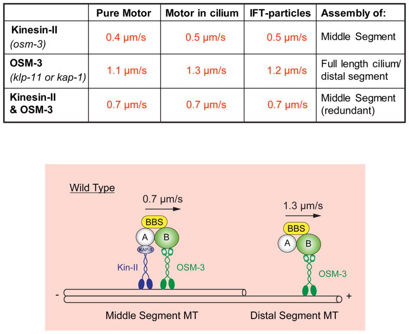
Figure 4.
Model for IFT along C. elegans sensory cilia inferred from in vitro motility and in vivo transport assays. Upper table shows rates of MT gliding driven by purified recombinant kinesin-II, OSM-3, or mixtures of kinesin-II and OSM-3 that reconstitute the rates of transport of IFT-particles along cilia driven by kinesin-II alone (in osm-3 or bbs mutants), OSM-3 alone (in kinesin-II or bbs mutants) or kinesin-II/OSM-3 together (along wild type middle segments). Lower panel, cartoon model showing how kinesin-II and OSM-3 together move IFT particles along the cilium middle segments to build the middle segments, then OSM-3 alone moves IFT-particles along the distal segments to assemble the distal segments. See (Ou et al., 2005; Pan et al., 2006; Snow et al., 2004).

