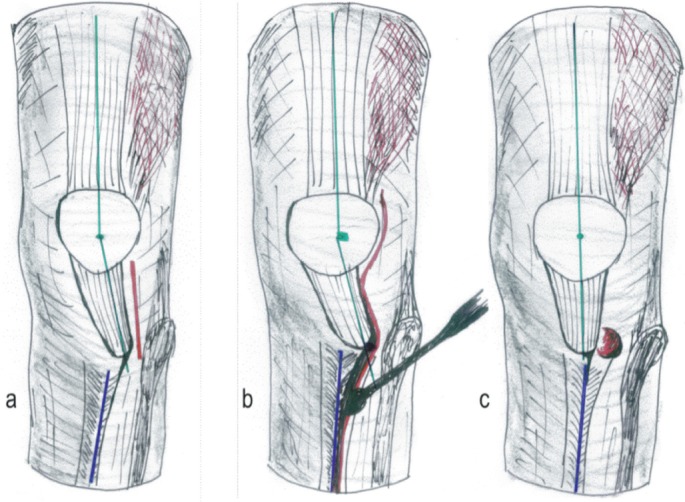
Figure 2.

Schematic drawing of left knee. a. Following arthroscopy, a lateral incision was made starting at the mid-patellar level and extending distally to include the anterolateral arthroscopy portal to reach 1 cm below the tibial tuberosity. b. and c. In cases with concomitant osteochondral fragments, the incision was extended proximally allowing access to the lateral femoral condyle or the patella facet (red). Starting at the tibial tuberosity, the periosteum was split along the tibial crest for 6–8 cm distally without opening the fascia of the tibialis anterior muscle (red). The periosteum was subsequently detached from the tibial crest with a large rasp (blue). In knee flexion, the patella tendon then slides spontaneously medially to track within the femoral groove. Patella alignment was restored (green).
