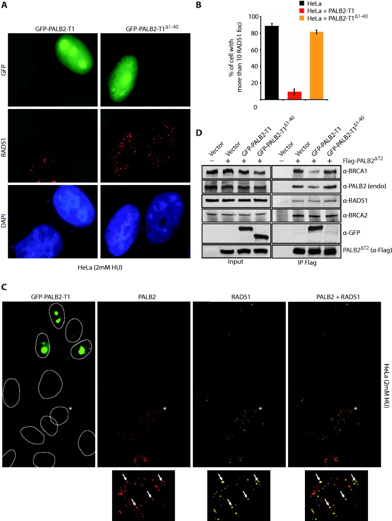
Figure 4.
Inhibition of RAD51 foci formation by the PALB2 coiled-coil domain. (A) Expression in Hela cells of GFP-PALB2-T1 with or without the coiled-coil domain (GFP-PALB2-T1Δ1–40). Immunofluorescence pictures with anti-GFP and anti-RAD51 are shown. (B) Quantification of the experiment shown in (A). (C) Expression in Hela cells of GFP-PALB2-T1 followed by immunofluorescence with anti-PALB2 (in red) and anti-RAD51 (in Far-red). The dotted lines delimit the DAPI staining. The cell indicated with an asterisk was enlarged at the bottom of the figure and arrows indicate a co-localization between PALB2 and RAD51 foci. (D) HEK293T cells were transfected with a vector alone or Flag-PALB2ΔT2, with GFP-PALB2-T1 or GFP-PALB2-T1Δ1–40. Flag-IPs were conducted, followed by western blotting with the indicated antibodies.

