Figure 1.

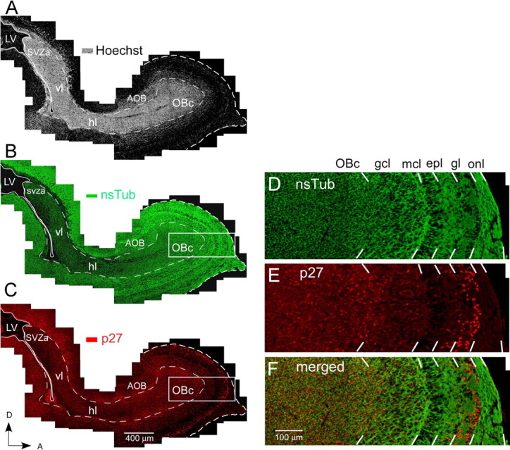
Overview of p27 expression in the WT mouse at P1 in the RMS and neuronal cell layers of the olfactory bulb. A–C, Identical parasagittal section stained with Hoechst 33342 (A), anti-nsTub (B), and anti-p27 (C). A, Bright-field photomicrograph of a parasagittal forebrain section stained with the nuclear dye Hoechst 33342 showing intense labeling of the RMS, which contains a high cell density compared with the surrounding brain regions. The four regions of the RMS labeled along its caudal–rostral axis are as follows: (1) the SVZa, an enlarged region of the SVZ overlying the anterior horn of the lateral ventricle; (2) the vl, a short segment of the RMS that descends ventrally from the SVZa; (3) the hl, the rostral extension of the RMS; and (4) the OBc, the terminal portion of the RMS in the center of the olfactory bulb. B, Fluorescent photomicrograph of the same section illustrated in A showing the expression pattern of nsTub in the RMS and neuronal cell layers of the olfactory bulb. A large percentage of cells in the RMS and neuronal cell layers of the olfactory bulb express nsTub, a marker for immature neurons. Most cells within the RMS are believed to be neuronal-restricted progenitor cells (Menezes and Luskin, 1994; Menezes et al., 1995; Law et al., 1999; Temple and Alvarez-Buylla, 1999; Rochefort et al., 2002; Falls and Luskin, 2005). While migrating, these progenitors express proteins characteristic of immature neurons, and many remain mitotically active until they reach their postmigratory destination. C, Fluorescent photomicrograph of the same section illustrated in A and B showing the expression pattern of p27 in the RMS and neuronal cell layers of the olfactory bulb. p27 is expressed throughout the RMS, particularly in the OBc. D–F, High magnification of the region outlined by a box in B and C. D, Anti-nsTub staining. E, Anti-p27 staining. F, Merged image of D and E. p27 is highly expressed in the OBc and in neuronal cell layers of the olfactory bulb (i.e., gcl and gl), where the neuronal cell marker nsTub also displays high expression levels. Scale bars: A–C, 400 μm; D–F, 100 μm. AOB, Accessory olfactory bulb; epl, external plexiform layer; mcl, mitral cell layer; OB, olfactory bulb; onl, olfactory nerve layer; LV, lateral ventricle; D, dorsal; A, anterior.
