
Fig. 2.
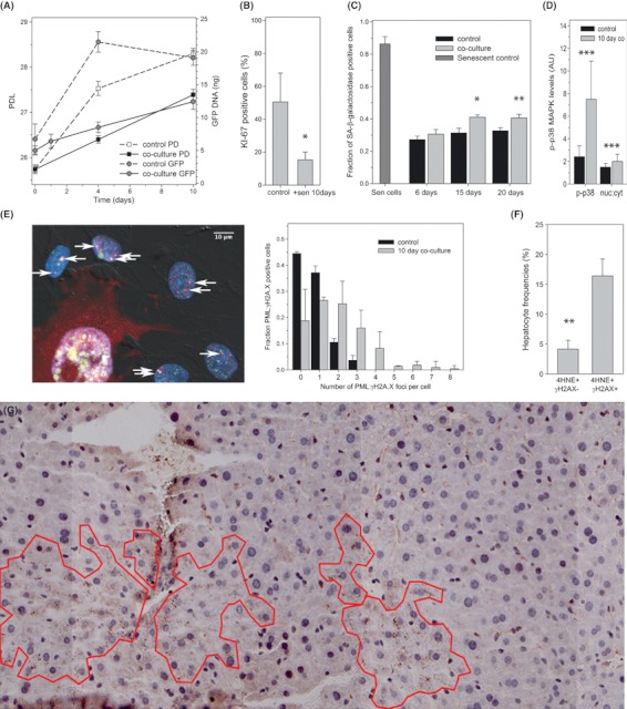
Senescent cells induce senescence in surrounding cells. (A) Impact of co-culture with senescent MRC5 cells on growth of MRC5-AcGFP-53BP1 cells measured by either cell counts (square symbols) or GFP-qPCR (circles). Cells were not replated during the experiment, leading to some density-mediated growth inhibition in control cells towards the end of the experiment. However, growth of bystander cells in co-culture was significantly slower (P < 0.01 at 4 and 10 days). (B) Frequencies of Ki67-positive MRC5-AcGFP-53BP1 cells after 10 day culture on their own (control) or 1:1 co-culture with senescent fibroblasts. Data are mean ± SEM, n = 4, P = 0.047. (C) Frequencies of Sen-β-Gal-positive MRC5-AcGFP-53BP1 bystander cells after the indicated time of co-culture with or without (control) senescent MRC5 cells. Senescent inducer cells were removed by blasticidin treatment for 6 days. Increases after 15 and 20 days co-culture are significant (anova with Tukey HSD P = 0.048 and 0.008, respectively). (D) Phospho-p38MAPK (P-p38) levels and nuclear:cytosolic ratios (measured by immunofluorescence) in MRC5-AcGFP-53BP1 after 10 days co-culture. Data are mean ± SD, P < 0.001 (T-test). (E) Co-localization (yellow) of γH2AX foci (green) with PML bodies (red) in bystander BJ-AcGFP-53BP1 cells after 10 day co-culture with senescent cells (marked by red RFP cytoplasmic fluorescence). Left: representative image; right frequency distributions of co-localization-positive cells. Means and distributions are significantly different (P < 0.0000, Mann–Whitney U-test). (F) Percentage of hepatocytes in adult mice liver showing cytoplasmic 4HNE staining together with (4HNE+ γH2AX+) or without (4HNE+ γH2AX−) nuclear DDR. Data are mean ± SE, n = 4 animals. (G) Representative tiled image of mouse liver (9 months old) immunostained for 4-HNE. Areas of clustered 4-HNE-positive cells are outlined. DDR, DNA damage response.