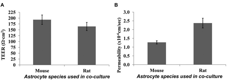
Figure 5.

Comparison of endothelial cell monolayers grown in the presence of mouse versus rat astrocytes seeded on the bottom of the well. (A) TEER data; and (B) sodium fluorescein permeability data. Endothelial cells were seeded at 4 × 10⁵ cells/cm2, astrocytes were seeded at 4 × 10⁴ cells /cm2 two days before endothelial cells were seeded, and all cultures were fed using media-feeding strategy 3. Data was collected on day 5 of endothelial cell growth on the membrane, and error bars show the standard deviation collected over three independent experiments.
