Figure 4.
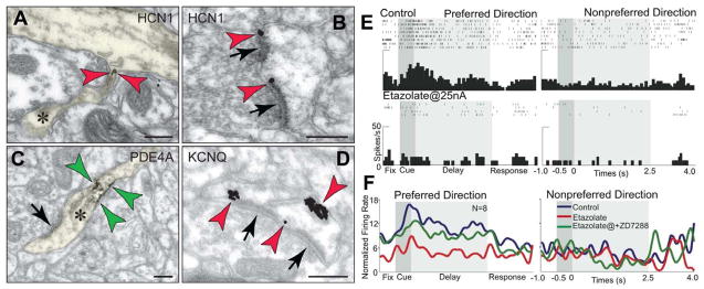
cAMP signaling in primate dlPFC differs from traditional neuroplasticity. A and B. Dendritic spines in monkey layer III dlPFC sequester cAMP-gated HCN channels (red arrowheads), positioned to translate cAMP “hot spots” to network connectivity patterns. HCN channels are found in the spine neck (A) and in the perisynaptic membranes flanking excitatory (network) synapses (B), positioned to gate impulses to the parent dendrite. C. PDE4A, which hydrolyzes cAMP and terminates its actions, presents an identical expression pattern in the neck region (green arrowheads); please see also Figure 3A. A–C adapted from (Paspalas et al., 2012). D. KCNQ potassium channels are also expressed in dlPFC spines, and are shown here in perisynaptic and extrasynaptic membranes (red arrowheads). Asterisks mark spine apparata; arrows point to excitatory-like synapses. Scale bars, 100 nm. E. Increased cAMP signaling reduces dlPFC Delay cell firing. Iontophoresis of the PDE4 inhibitor, etazolate (25 nA), onto a dlPFC Delay cell rapidly reduces task-related firing in a monkey performing a working memory task. F. Population response of 8 Delay cells with delay-related firing under control conditions (blue trace), that is markedly reduced by iontophoresis of etazolate (red trace). Blockade of HCN channels with co-iontophoresis of ZD7288 (10 nA, green trace) restores normal firing patterns, demonstrating physiological interactions between HCN channels and cAMP signaling in the primate dlPFC. Adapted from (Wang et al., 2007).

