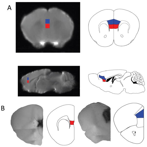
Figure 2. IL and PL MRI seeds for DTI tractrography and PL BDI injection site.
Each brain is placed in a standard space that is also matched (same dimensions, with the ability to overlay the same rulers place on the atlas, to stereotactically determine regions of interest) to the Paxinos and Franklin Mouse Atlas used to determine areas of connectivity from the IL (shown in red) and Pl (shown in blue) seeds. A). Presented is a representation of the MRI images matched to slices in the atlas, as determined by specific anatomical landmarks (particularly the white matter tracts of the corpus callosum, the lateral and third ventricles and the aqueduct to the 4th ventricle, the internal capsule, and the hippocampus). B) Shows the injection site of the BDA/ABC/DAB in the PL and IL area at 4x magnification using phase contrast. The representative atlas slice is shown to the right with IL highlighted in red and Pl highlighted in blue, for the PL injection the cingulate outlined more dorsally and the medial orbital cortex outlined below.

