
Figure 3 .
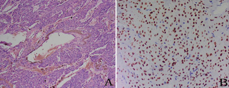
Pulmonary sclerosing hemangioma was confirmed by surgical resection and postoperative pathological diagnosis. (A) Two of the typical histological characteristics: hemorrhagic and papillary areas (×100). (B) Both the cuboidal surface cells and polygonal cells showed thyroid nuclear factor 1 staining (×200).