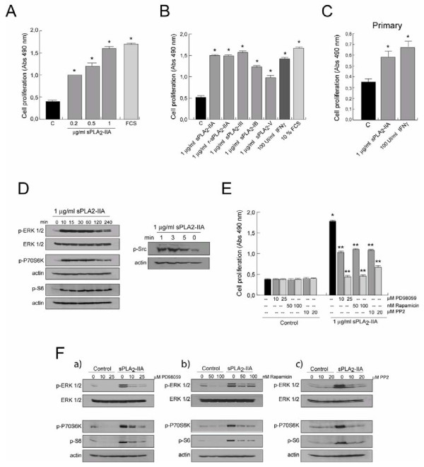
Figure 1 .
sPLA2-IIA modulates proliferation in microglial cells. BV-2 cells were stimulated with different doses of sPLA2-IIA and 10% of FCS (A), or with the indicated stimuli (B). Primary microglial cells were stimulated with 1 μg/ml of sPLA2-IIA or 100 UI/ml of IFNγ (C). After 24 h of incubation at 37°C, cell proliferation was investigated and expressed as optical density (OD) values ± SD. Values are the average of three separate experiments in quadruplicate (*P < 0.001 vs. control cells). (D) BV-2 microglial cells were incubated with sPLA2-IIA for different times. Cell lysates were collected and subjected to western blot analysis using Abs against p-Src, ERK 1/2, p-ERK1/2, p-P70S6K, p-rS6 and actin. (E) BV-2 cells were treated at 37°C for 30 minutes in the presence or absence of the indicated inhibitors, and then were stimulated with 1 μg/ml of sPLA2-IIA. After 24 h of incubation, cell proliferation was investigated and expressed as OD values ± SD. Values are the average of three separate experiments in quadruplicate (*P < 0.001 vs. control cells, **P < 0.001 vs. sPLA2-IIA-treated cells). (F) BV-2 cells were treated as in (D). After 15 minutes stimulation, whole cell lysates were extracted and protein phosphorylation was assessed by western blotting using p-ERK, p-P70S6 and p-rS6 antibodies. Membranes were always stained with Ponceau S as a loading control.

