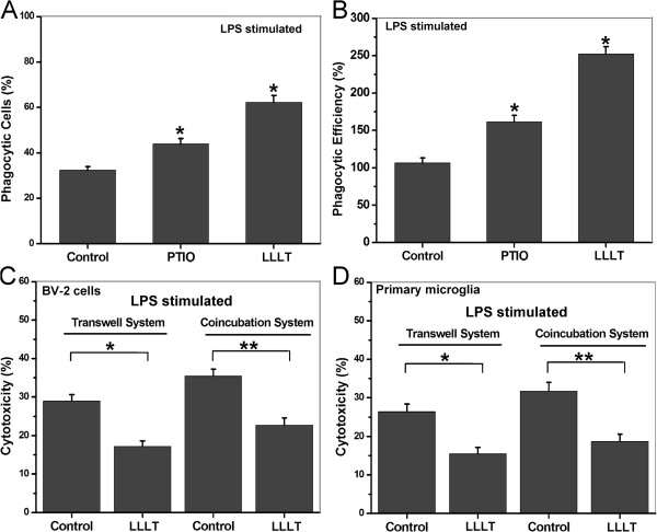
Figure 7.
Correlations between inflammatory mediators and the phagocytic response induced by LLLT in LPS-activated microglial. (A and B) LPS-activated BV-2 cells pretreated with or without 200 μM carboxy-PTIO. Then the cells with or without LLLT (20 J/cm2) treatment were incubated with microspheres for 30 min. (A) Quantification of LLLT-mediated microglial phagocytosis. (B) Number of microspheres taken up per BV-2 cell. (n = 4; *P <0.05 versus control cells). (C and D) SH-SY5Y cells cultured with LPS-activated BV-2 cells or primary microglia (at an E:T ratio of 8:1) in a Transwell™ cell-culture system or directly in the mixed co-culture system for 24 h. After incubation, target cells that were doubly positive for CFSE and PI were analyzed by flow cytometry (n = 4; *P <0.05 and **P <0.05 versus indicated cells). CFSE, carboxyfluorescein diacetate succinimidyl ester; LLLT, low-level laser therapy; LPS, lipopolysaccharide; PI, propidium iodide.

