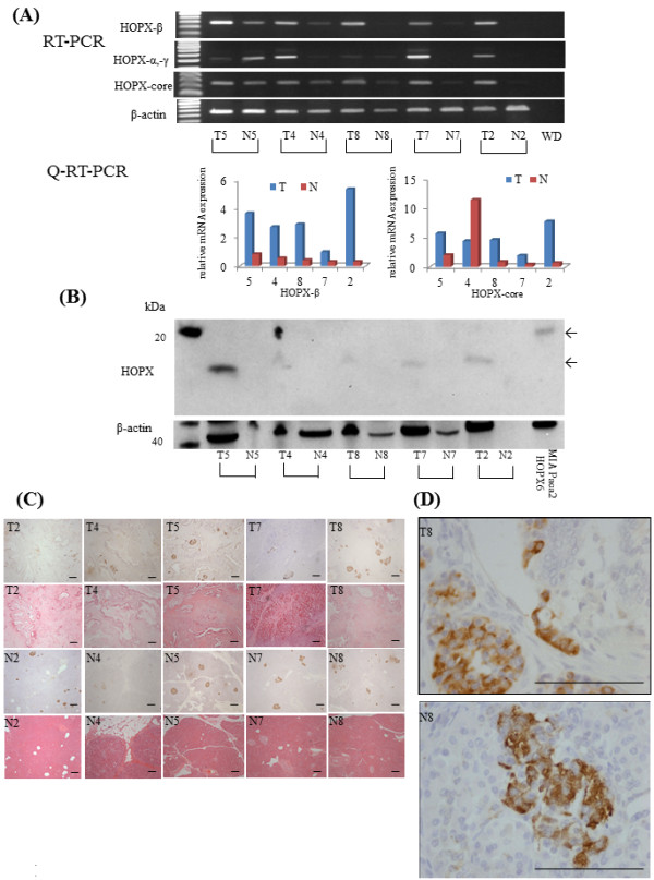
Figure 3.

HOPX expression status in PC. (A) Expression level of HOPX in PC was tested in RT-PCR (top panel) and Q-RT-PCR (bottom panel). T, primary tumor; N, corresponding pancreatic tissue. (B) Expression level of HOPX in PC was examined by western blotting. (C) Immunohistochemical staining for HOPX in primary tumor (top panel) and normal tissue (bottom panel), with hematoxylin eosin staining (original magnification, X40). These immunohistochemical stainings were performed by short term exposure of DAB. (D) In this condition, islet cells only stained (original magnification, X400). scale Bars, 100 μm.
