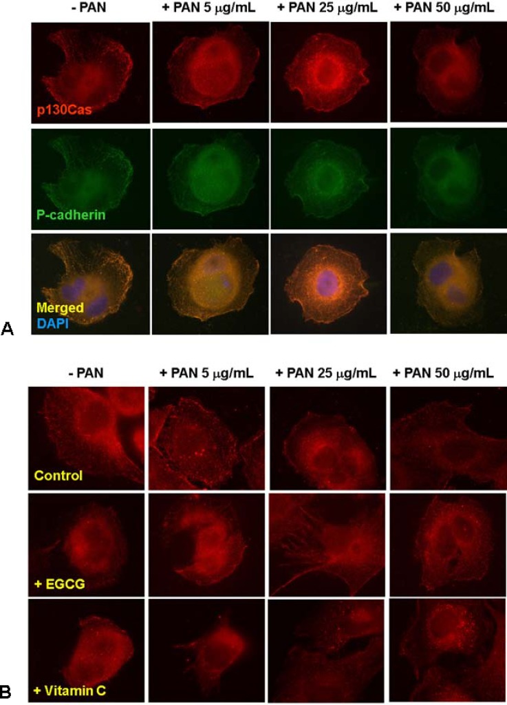
Fig. 1.
Distributional changes of p130Cas. p130Cas showed a diffuse cytoplasmic distribution with accumulation at distinct peripheral areas visible as short stripes in physiologic condition and colocalized with P-cadherin. The fluorescences of p130Cas protein were internalized and became granularly by PAN in a dose-dependent manner (A, rat glomerular epithelial cells). Such pathologic changes were reversed by EGCG and vitamin C (B, mouse podocyte). Magnification, ×400.

