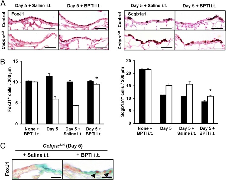
Figure 7.
Intratracheal treatment with BPTI restored FoxJ1+ cells in vivo. (A) Immunohistochemistry of FoxJ1 and Scgb1a1 in the terminal bronchioles is shown on Day 5 after naphthalene injury, with or without daily BPTI treatment. FoxJ1+ cells were increased after BPTI treatment in the CebpαΔ/Δ mice to a level similar to that in control mice. Scale bars, 50 μm. (B) The numbers of stained FoxJ1 and Scgb1a1 cells lining 200 μm of the terminal bronchiolar epithelium from the bronchoalveolar duct junctions were quantitated. The number of FoxJ1+ cells in the terminal bronchioles of the CebpαΔ/Δ mice was restored by BPTI treatment. The intratracheal injection of BPTI into noninjured mice (None + BPTI i.t.) did not influence the number of FoxJ1+ and Scgb1a1+ cells. In CebpαΔ/Δ mice, the proportion of Scgb1a1-stained cells decreased after an intratracheal injection of BPTI, consistent with the increase in FoxJ1+-stained cells. *P < 0.05 versus Day 5 + Saline, according to ANOVA (n = 4/group). (C) Intratracheal treatment with BPTI increased the double-positive (FoxJ1, red; Lac Z, green) cells in the terminal bronchioles of Rosa–CebpαΔ/Δ mice, supporting the concept that BPTI restored the regeneration of ciliated cells in Rosa–CebpαΔ/Δ mice. Scale bars, 20 μm (n = 3/group).

