Figure 6.
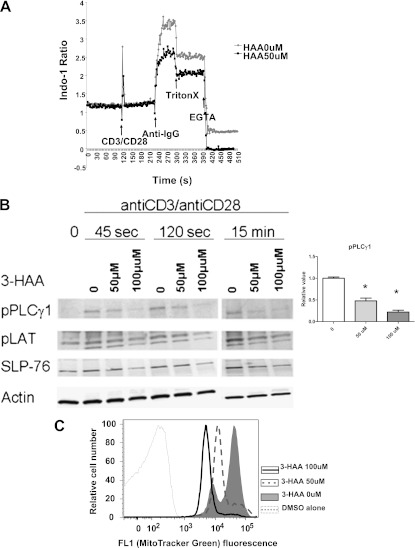
IDO and 3HAA inhibit TCR-mediated Ca2+ mobilization, signaling molecules, and mitochondrial content. (A) A representative example (n = 5) of decreased Ca2+ influx in T cells after TCR (anti-CD3/anti-CD28) activation in the presence of 3HAA (50 μM), as measured by indo-1 staining. Arrows indicate the timing of cell activation with respect to Ca2+ measurements. (B) Immunoblots of lysates from naive C57BL/6 cells stimulated with anti-CD3/anti-CD28 activation after 45 seconds, 120 seconds, and 15 minutes, with and without 3HAA exposure. A representative analysis (n = 3 independent experiments) is illustrated for phosphorylated (p) PLCγ1, pLAT, and SLP-76, with β-actin as internal loading control. The graph illustrates the relative pPLCγ1 concentration at 120 seconds for untreated cells and 3HAA treatment. (C) Decreased mitochondrial content of CD4+ and CD8+ T cells was measured by MitoTracker Green staining. Splenocytes cultured with the indicated concentrations of 3HAA were stained with anti-CD4 and anti-CD8, as well as 75 nM MitoTracker Green, for 20 minutes at 37°C. Splenocytes were then washed and their fluorescence was determined by flow cytometry. The fluorescence histogram is representative of three independent experiments.

