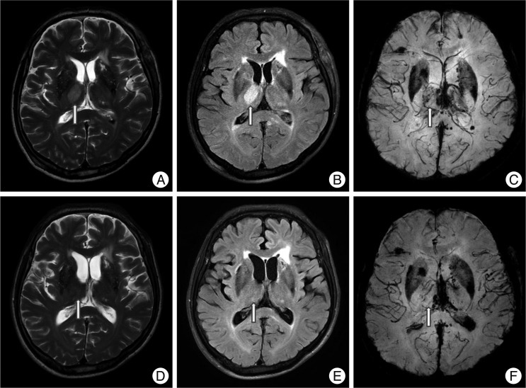
Fig. 2.
A non-hemorrhagic lesion of the thalamus area. A 57-year-old male was admitted due to a pedestrian car accident, with a Glasgow coma scale of 7 on admission. MRI shows a non-hemorrhagic lesion at the right thalamus (arrow). Non-hemorrhagic lesions show high signal intensity at T2W1 (A) and FLAIR (B), but a dark signal cannot be seen on SWI (C). However, at one month follow-up, the previous high signal at T2W1 (D), FLAIR (E) is not seen and SWI (F) still showed no dark signals.

