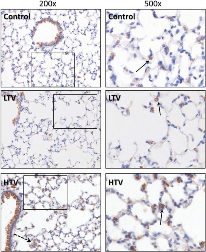
Figure 4.
COX-2 expression in murine lungs. Murine lungs were formalin-fixed in situ and immunohistochemically stained with antibodies directed against COX-2. In addition to the expected prominent staining of bronchiolar epithelium in all groups, significant staining of mononuclear cells was evident, showing granular cytoplasmic positivity to COX-2. These cells were confirmed to be of the monocyte/macrophage lineage by costaining for CD45 and CD68 (not shown). Note the presence of COX-2–positive alveolar macrophages in the HTV group (dashed arrow), in addition to interstitial monocytes and macrophages located in close proximity to the alveolar space (solid arrows). Images represent 5-μm histologic sections. Boxed areas in left panels are presented at ×500 magnification in right panels.

