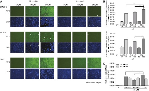
Figure 2.
CDC promotes the association of curcumin with lung epithelial cells. Calu-3 cell monolayers on Transwell inserts were exposed to CDC, DMSO-C, or EtOH-C in either the apical (AP→BL) or basolateral (BL→AP) compartments for 180 minutes, with shaking at 37°C in a 5% CO2 incubator. (A) The inserts were then washed and incubated for another 30 minutes with the nucleus-specific dye Hoechst 33342 (10 μg/ml) in the apical compartment, and with dye-free HBSS transport buffer in the basolateral compartment. Cell monolayers were examined with a Nikon TE 2000 fluorescence microscope (×10 objective) (Nikon Instruments, Melville, NY). Curcumin was visualized in the FITC channel, and nuclei were visualized in the DAPI channel. Regions where cells have detached or where monolayer integrity is visibly compromised are indicated with an asterisk. Scale bar of 80 μm is displayed. Representative images are shown from three independent experiments. (B and C) After exposure, cells were washed, and cell-associated curcumin was extracted with 1% Triton X-100. Extracted curcumin was quantitated (mass per cell) by fluorescence, using a plate reader. Amounts of the paracellular marker LY are also shown. (B) Cell-associated curcumin as a function of the initial donor concentration for AP→BL (top) and BL→AP (bottom) transport. (C) Cell-associated curcumin as a function of transport direction at 50 μM (n = 6). **P < 0.01. ***P < 0.001.

