Figure 6.
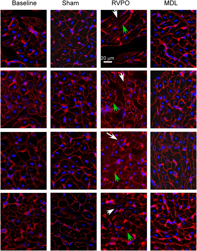
Talin. Representative photomicrographs were obtained from 16-μm transverse sections of the RV free wall in pigs before (Baseline) and after 2 hours of acute RVPO immunostained for talin (red) and with (2-(4-amidinophenyl)-1H-indole-6-carboxamidine) (DAPI) (in blue, for nuclei). The second biopsy was obtained from a location at least 2 cm away from the first biopsy, and in a more proximal perfusion territory, to avoid sampling a region potentially affected by the first biopsy. Under baseline conditions, the talin signal was largely localized to the cell surface. After 2 hours of acute RVPO, patchy loss of the regular signal (white arrows) and areas of blurring (green arrows) were evident. RV myocardium from pigs identically instrumented and subjected to 2 hours of anesthesia without RV pressure overload (Sham) did not exhibit any significant differences from baseline biopsies, indicating that 2 hours of anesthesia without RVPO did not cause changes in talin organization according to immunohistology. Biopsies from pigs subjected to 2 hours of RVPO but pretreated with MDL-28170 (MDL) exhibited structures more like those of Baseline and Sham than RVPO. Image sets were obtained on a Leica digital deconvolution microscope (Leica Microsystems, Buffalo Grove, IL), using a ×63 oil immersion objective (NA 1.4) with 0.5-μm z-dimension steps, and processed using constrained iterative deconvolution. Image intensity and contrast were equalized among all images. Scale bar = 20 μm.

