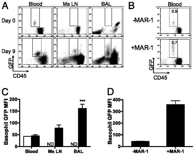
Figure 3. Basophil GFP MFI in tissues.
(A) FACs plots of non-CD4 cells from uninfected, day 9 blood, BAL and Ms LN of primary Nb infected G4/G4 mice, showing FcεR1α+GFP+ basophils. (B) FACS plot of peripheral blood 12hr following i.v. administration of MAR-1. Indicated percentages represent the proportion of GFP+ basophils among total CD45+ cells. (C) MFI of basophils present in blood, BAL and mesenteric lymph nodes at days 0 (clear) and 9 (black) post infection. (D) MFI of basophils present in peripheral blood at day 1 ± MAR-1. Data points shown indicate mean ± SE from three individual animals from two experiments. ***, P ≤ 0.0001; **, P ≤ 0.001; *, P ≤ 0.01; no asterisk, P > 0.05, relative to day 9 blood basophils with Student’s t-test. ND, None Detected.

