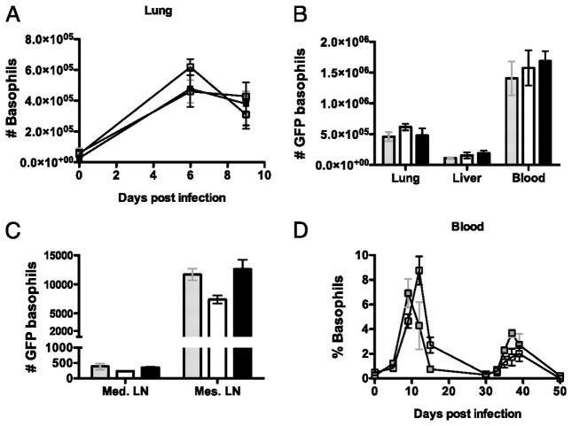
Figure 4. The number of GFP producing basophils in response to a secondary Nb infection is not dependent on IL-4 or STAT6.
The total number of GFP expressing basophils were detected following a secondary Nb infection in G4/IL-4 (grey), G4/G4 (clear) and G4/G4 xSTAT6 ko (black) mice. (A) Kinetics of GFP basophil induction in the lung. (B) Total number of GFP basophils in the lung, liver, blood, (C) mediastinal lymph node and mesenteric lymph node day 6 after infection. (D) Kinetics of GFP basophil induction in the BAL following primary (day 0) and secondary (Day 30) infection with Nb in G4/IL-4xSTAT6 ko (grey) and G4/G4xSTAT6 ko (black) mice. Data points shown indicate mean ± SE from three individual animals from two experiments.

