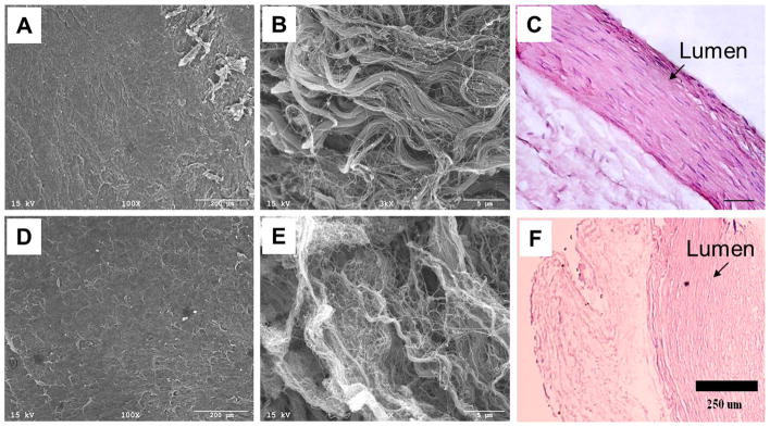
Figure 5. SEM & H&E of the HUA.
(A,B,C) The scanning electron micrograph and hematoxylin and eosin staining of the human umbilical artery before decellularization show a more orderly structure of fibers. (D,E,F) The decellularized human umbilical arterial fibers have been unwoven and now show a more loosely-packed, slightly randomized structure.

