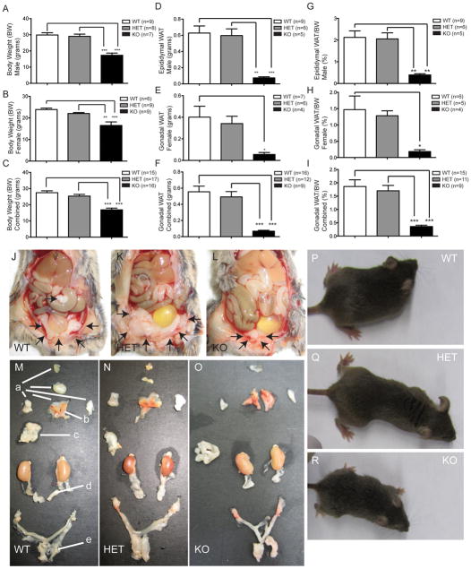
Figure 1. Body weight and gonadal WAT are reduced in VGF knockout mice.
Body weights (grams) of male and female Vgf−/Vgf−, Vgf+/Vgf−, and Vgf+/Vgf+ mice were measured at 9 weeks of age (panels A–C); male and female Vgf−/Vgf− mice had significantly lower body weights compared to Vgf+/Vgf−, and Vgf+/Vgf+ mice. In panel D, gonadal fat pad weights (grams) were significantly reduced in Vgf−/Vgf− (n=5 male, 4 female), compared to Vgf+/Vgf− (n=6 male, 7 female) and Vgf+/Vgf+ (n=9 male, 7 female). No significant differences in body weight, fat pad weight, or the ratio of fat pad weight to body weight, between Vgf+/Vgf− and Vgf+/Vgf+ mice, were noted (panels A–I). Body weights and epididymal and ovarian fat pad weights were measured for each genotype at 9–10 weeks of age; values are mean ± SEM, *p = 0.0430, **p = 0.0031, ***p < 0.0001 compared to wild type (ANOVA and Tukey’s multiples comparison). Eight week old female mice [Vgf+/Vgf+ WT (24.6 grams body weight), Vgf+/Vgf− HET (20.5 grams), and Vgf−/Vgf− KO (13.6 grams)] (panels P–R, respectively) were transcardially perfused with 4% paraformaldehyde in PBS, intraperitoneal fat depots were exposed (panels J–L), and the subcutaneous and visceral depots were dissected and placed on a mouse template to show their location in the body (panels M–O). Fat depots are labeled with lower case letters: a (subcutaneous anterior: deep & superficial cervical, interscapular, subscapular, axillo-thoracic); b (interscapular brown adipose tissue); c (mesenteric); d (perirenal, periovarian, parametrial, perivesical); e (subcutaneous posterior: dorsolumbar, inguinal, gluteal) (Murano, et al. 2009).

