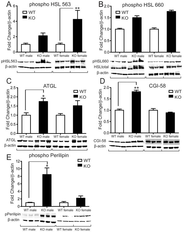
Figure 2. Altered expression of proteins involved in lipolysis in VGF knockout compared to wild type WAT.
WAT tissues were collected from Vgf−/Vgf− knockout mice (n=11–13) and Vgf+/Vgf+ wild type littermate controls (n=9–11), and protein expression was determined using western blot analysis and Image J as described in Materials and Methods. Values are expressed as fold change in knockout relative to wild type WAT, normalized to β-actin as a loading control, and statistical significance was determined using the two-tailed Student’s t test (*p < 0.05). Levels of phospho-HSL (ser563) were significantly increased in VGF KO WAT (panel A, *p = 0.0012), while phospho-HSL (ser660) levels were unchanged (panel B). Total HSL levels were also unchanged (see Table 1). Expression of ATGL (panel C, *p = 0.0312), CGI-58 (panel D, *p = 0.0074), and phospho-perilipin (panel E, *p = 0.0317) were significantly increased in VGF knockout WAT.

