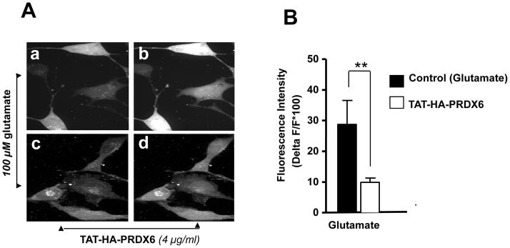
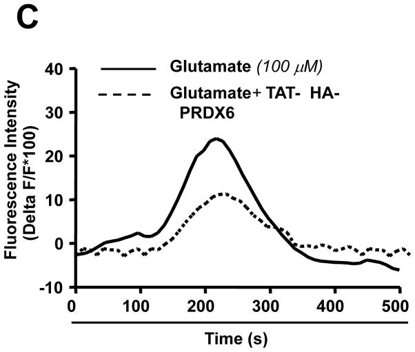
Figure 10.
PRDX6 inhibits Ca2+ influx into RGC-5 following glutamate treatment. Intracellular Ca2+ level was measured using the cell permeable Fluo-4 AM, fluorescent calcium indicator dye. Cells were cultured on cover slips in DMEM medium with TAT-HA-PRDX6 or mutant PRDX6 protein followed by glutamate treatment. (A) Fluorescent images were taken before and during application of 100 μM glutamate without (a, b) or with 4 μg/ml TAT-HA-PRDX6 (c, d). Pretreatment with TAT-HA-PRDX6 significantly reduced the increase in Fluo-4 fluorescence produced by application of 100 μM glutamate indicating TAT-HA-PRDX6 controls the glutamate-induced increase in Ca2+. (B) Histogram showing fluorescent intensity of the cells treated with glutamate in presence (empty bar, **p <0.001 vs. control) or absence of TAT-HA-PRDX6 (black bar). (C) Graph showing fluorescent intensity of cells at different time points (seconds).


