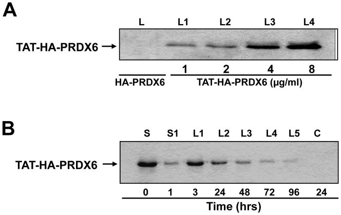
Figure 5.
(A) Concentration dependent transduction of TAT-HA-PRDX6. Recombinant protein was added to culture media and transduction of TAT-HA-PRDX6 was assessed after 24 hr. Cells were washed, protein was extracted and used for Western analysis using anti-His HRP antibody. Lanes, L1–L4: cell lysates from the cells cultured with 1, 2, 4, 8 μg/ml protein, and lane L denotes the lysate extracted from cells cultured with HA-PRDX6 only. (B) Western analysis showing stability of TAT-HA-PRDX6 transduced into RGCs. Lane, S: culture media just after addition of recombinant protein (0hr); lane, S1: Culture supernatant after 1 hr; Lanes, L1 to L5: Cell lysate after 1, 3, 24, 48, 72 and 96 hrs; C: Control (HA-PRDX6 without TAT). Results revealed the intracellular transduction of TAT-HA-PRDX6 where as (HA)-PRDX6 with flag tag (HA) only could not internalize into cells (lane C).

