
Figure 1.
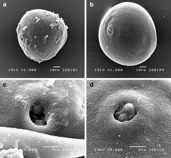
Comparison of the (A, C) A. mellifera harvested bee pollen with that of (B, D) corn pollen by SEM showing (A, B) the external morphology (1000 x magnification) and (C, D) the germination pore and ornamentation of the pollen exine (5000 x magnification). Micrographs shown are representative of those seen from 10 independent pollen grains.